superimposing structures with Chimera

On Feb 10, 2022, at 1:42 PM, Kaina Quintero Chavez <kaina.quintero@uabc.edu.mx> wrote:
Hi, My name is Kaina. I am a current college student in UABC. I am currently doing my thesis in biology and came upon using UCSF chimera and am in a stuck position on how to use the superimpose feature. I was hoping you can guide me or reference me to a workshop/ course? anything really to get the job done. Thanks, hope to here from you. -- Kaina
Hi Kaina, We usually recommend the chimera-users@cgl.ucsf.edu address for asking questions -- I CC'd it here, hope that's OK. Also it would be helpful to include more information when you send a question, like what you are trying to superimpose, what you tried already, what problem(s) caused you to be stuck, etc... but I will try to answer below. There are lots of different ways to superimpose structures in Chimera and it depends on your situation as to which one you would use. There is a discussion of the different ways here: <https://www.rbvi.ucsf.edu/chimera/docs/UsersGuide/superposition.html> If you have atomic structures of proteins or nucleic acid chains, probably the easiest approach is to try using the Matchmaker tool, menu: Tools... Structure Comparison... Matchmaker. You would choose one structure as the reference and the other one as the one to match, and then just click "Apply" to see if the default settings work. Here is the help for Matchmaker <https://www.rbvi.ucsf.edu/chimera/docs/ContributedSoftware/matchmaker/matchm...> ...and this tutorial includes examples of using it <https://www.rbvi.ucsf.edu/chimera/docs/UsersGuide/tutorials/alignments.html> There is also a "matchmaker" command that does the same thing, and the help includes some example commands: <https://www.rbvi.ucsf.edu/chimera/docs/UsersGuide/midas/mmaker.html> However, maybe your situation is different and you would need to use one of the other methods discussed in the first link above. I hope this helps, Elaine ----- Elaine C. Meng, Ph.D. UCSF Chimera(X) team Department of Pharmaceutical Chemistry University of California, San Francisco

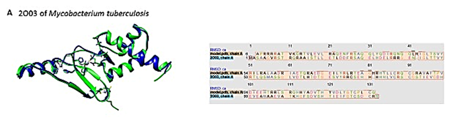
Hi, thank you very much for the information, and I apologize for not being so detailed about my question/problem. With the information provided I was able to superimpose my two proteins but still not in the way I want to. I was given a photo of how it should look and my structure just does not seem to orientate the same way using the default method. I hope it is not too much trouble and are able to help me out. I attached my two proteins and a copy of the picture I was given to replicate. this with the objective to be able to duplicate the work with more complex proteins. [image: image.png] ps. I am sorry for the quality of the photo, this was the best I could sharpen since they only have this one copy and are unable to duplicate the work. [image: image.png] As you can see I am still left with some part of the protein not overlapped and my question and inquiry is how can I modify the structure so both proteins overlap or if the program can make the torshions or necessary turns so they fit.
Hi Kaina, The problem is not how to superimpose the structures, which you are already doing correctly. The problem is that the structures are in a totally different conformation from each other. Also, your "map" PDB is a dimer of two copies of the protein as chains A and B, whereas 2o03 is a monomer with just chain A. Even if I hide or delete one of the chains in the "map" PDB, however, it is clearly a very different conformation than the 2o03 monomer, so that is the main problem. However, you can't just start changing torsions in a 3D structure and expect that structure to be valid... and it would be very difficult to do by hand, perhaps impossible. In my opinion, the only way (without fairly advanced modeling) to get something that looks like your first image is to already have another monomer structure that is in a similar conformation to 2o03. This image shows 2o03 (tan color) in similar orientation to your first image, superimposed with your "map" PDB chain A in transparent blue. The other monomer of the "map" PDB is in transparent pink. About half of the blue chain matches well, but then a difference in conformation in the middle of the blue chain sends the rest of that chain in a different direction to what is in 2o03. If you're just making a schematic and you don't care if the protein structure is really valid, you could delete one chain of your "map" structure and try rotating the backbone phi,psi angles in the middle of the other chain. You'd probably want to hide ribbon and show backbone atoms first. However, I would not want to try that myself, because it is usually much harder than you think to make the structure look the way you want. I hope this makes sense, Elaine ----- Elaine C. Meng, Ph.D. UCSF Chimera(X) team Department of Pharmaceutical Chemistry University of California, San Francisco
On Feb 11, 2022, at 11:50 AM, Kaina Quintero Chavez via Chimera-users <chimera-users@cgl.ucsf.edu> wrote:
Hi, thank you very much for the information, and I apologize for not being so detailed about my question/problem. With the information provided I was able to superimpose my two proteins but still not in the way I want to. I was given a photo of how it should look and my structure just does not seem to orientate the same way using the default method. I hope it is not too much trouble and are able to help me out. I attached my two proteins and a copy of the picture I was given to replicate. this with the objective to be able to duplicate the work with more complex proteins. <image.png> ps. I am sorry for the quality of the photo, this was the best I could sharpen since they only have this one copy and are unable to duplicate the work.
<image.png> As you can see I am still left with some part of the protein not overlapped and my question and inquiry is how can I modify the structure so both proteins overlap or if the program can make the torshions or necessary turns so they fit. <map3773c.pdb><2o03.pdb>_______________________________________________ Chimera-users mailing list: Chimera-users@cgl.ucsf.edu Manage subscription: https://www.rbvi.ucsf.edu/mailman/listinfo/chimera-users
Also keep in mind that usually the reason to make a figure showing a superposition is to demonstrate real similarities and differences between structures. If you manually alter one structure to make it look like the other one, it seems like that would defeat the purpose of making such a figure. Elaine
On Feb 11, 2022, at 1:30 PM, Elaine Meng via Chimera-users <chimera-users@cgl.ucsf.edu> wrote:
Hi Kaina, The problem is not how to superimpose the structures, which you are already doing correctly. The problem is that the structures are in a totally different conformation from each other. Also, your "map" PDB is a dimer of two copies of the protein as chains A and B, whereas 2o03 is a monomer with just chain A. Even if I hide or delete one of the chains in the "map" PDB, however, it is clearly a very different conformation than the 2o03 monomer, so that is the main problem.
However, you can't just start changing torsions in a 3D structure and expect that structure to be valid... and it would be very difficult to do by hand, perhaps impossible. In my opinion, the only way (without fairly advanced modeling) to get something that looks like your first image is to already have another monomer structure that is in a similar conformation to 2o03.
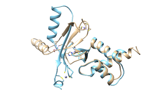
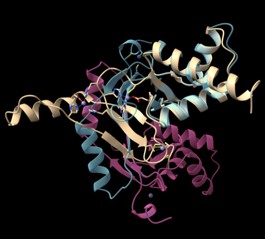
This image shows 2o03 (tan color) in similar orientation to your first image, superimposed with your "map" PDB chain A in transparent blue. The other monomer of the "map" PDB is in transparent pink. About half of the blue chain matches well, but then a difference in conformation in the middle of the blue chain sends the rest of that chain in a different direction to what is in 2o03.
<kaina.png>
If you're just making a schematic and you don't care if the protein structure is really valid, you could delete one chain of your "map" structure and try rotating the backbone phi,psi angles in the middle of the other chain. You'd probably want to hide ribbon and show backbone atoms first. However, I would not want to try that myself, because it is usually much harder than you think to make the structure look the way you want.
I hope this makes sense, Elaine ----- Elaine C. Meng, Ph.D. UCSF Chimera(X) team Department of Pharmaceutical Chemistry University of California, San Francisco
On Feb 11, 2022, at 11:50 AM, Kaina Quintero Chavez via Chimera-users <chimera-users@cgl.ucsf.edu> wrote:
Hi, thank you very much for the information, and I apologize for not being so detailed about my question/problem. With the information provided I was able to superimpose my two proteins but still not in the way I want to. I was given a photo of how it should look and my structure just does not seem to orientate the same way using the default method. I hope it is not too much trouble and are able to help me out. I attached my two proteins and a copy of the picture I was given to replicate. this with the objective to be able to duplicate the work with more complex proteins. <image.png> ps. I am sorry for the quality of the photo, this was the best I could sharpen since they only have this one copy and are unable to duplicate the work.
<image.png> As you can see I am still left with some part of the protein not overlapped and my question and inquiry is how can I modify the structure so both proteins overlap or if the program can make the torshions or necessary turns so they fit. <map3773c.pdb><2o03.pdb>_______________________________________________ Chimera-users mailing list: Chimera-users@cgl.ucsf.edu Manage subscription: https://www.rbvi.ucsf.edu/mailman/listinfo/chimera-users
_______________________________________________ Chimera-users mailing list: Chimera-users@cgl.ucsf.edu Manage subscription: https://www.rbvi.ucsf.edu/mailman/listinfo/chimera-users
Thanks, and correct I am making a schematic. 2o03 is a known protein and “map” is a hypothetical protein. I want to compare both by overlapping and read the difference in the zinc distance. For this I would be hiding chain A of “map” and overlapping chain B to 2o03. In the case of rotating the backbone, in this case “map” since it’s the hypothetical protein, I would have to know what residues are not superimposed select those, rotate and this would adjust to the 2o03 ? Or would I have to make the rotation similar to 2o03 and redo the matchmaker? Again thanks for the helpful insight, I did try and read through the manual but didn’t get nowhere. On Fri 11 Feb 2022 at 1:30 p.m. Elaine Meng <meng@cgl.ucsf.edu> wrote:
Hi Kaina, The problem is not how to superimpose the structures, which you are already doing correctly. The problem is that the structures are in a totally different conformation from each other. Also, your "map" PDB is a dimer of two copies of the protein as chains A and B, whereas 2o03 is a monomer with just chain A. Even if I hide or delete one of the chains in the "map" PDB, however, it is clearly a very different conformation than the 2o03 monomer, so that is the main problem.
However, you can't just start changing torsions in a 3D structure and expect that structure to be valid... and it would be very difficult to do by hand, perhaps impossible. In my opinion, the only way (without fairly advanced modeling) to get something that looks like your first image is to already have another monomer structure that is in a similar conformation to 2o03.
This image shows 2o03 (tan color) in similar orientation to your first image, superimposed with your "map" PDB chain A in transparent blue. The other monomer of the "map" PDB is in transparent pink. About half of the blue chain matches well, but then a difference in conformation in the middle of the blue chain sends the rest of that chain in a different direction to what is in 2o03.
If you're just making a schematic and you don't care if the protein structure is really valid, you could delete one chain of your "map" structure and try rotating the backbone phi,psi angles in the middle of the other chain. You'd probably want to hide ribbon and show backbone atoms first. However, I would not want to try that myself, because it is usually much harder than you think to make the structure look the way you want.
I hope this makes sense, Elaine ----- Elaine C. Meng, Ph.D. UCSF Chimera(X) team Department of Pharmaceutical Chemistry University of California, San Francisco
On Feb 11, 2022, at 11:50 AM, Kaina Quintero Chavez via Chimera-users < chimera-users@cgl.ucsf.edu> wrote:
Hi, thank you very much for the information, and I apologize for not being so detailed about my question/problem. With the information provided I was able to superimpose my two proteins but still not in the way I want to. I was given a photo of how it should look and my structure just does not seem to orientate the same way using the default method. I hope it is not too much trouble and are able to help me out. I attached my two proteins and a copy of the picture I was given to replicate. this with the objective to be able to duplicate the work with more complex proteins. <image.png> ps. I am sorry for the quality of the photo, this was the best I could sharpen since they only have this one copy and are unable to duplicate the work.
<image.png> As you can see I am still left with some part of the protein not overlapped and my question and inquiry is how can I modify the structure so both proteins overlap or if the program can make the torshions or necessary turns so they fit. <map3773c.pdb><2o03.pdb>_______________________________________________ Chimera-users mailing list: Chimera-users@cgl.ucsf.edu Manage subscription: https://www.rbvi.ucsf.edu/mailman/listinfo/chimera-users
-- Kaina
Hi Kaina, I don't think any of this makes sense. If you want to make a schematic with a protein conformation that looks like 2o03, just use the structure of 2o03. There is no reason to spend hours and hours trying to make your "map" protein look like 2o03 when you already have 2o03. Maybe my previous message made it sound doable to hand-rotate bonds to make your "map" protein conformation look like 2o03 but it's really not. Even as a Chimera expert, I don't think I could do it. I hope this clarifies the situation, Elaine ----- Elaine C. Meng, Ph.D. UCSF Chimera(X) team Department of Pharmaceutical Chemistry University of California, San Francisco
On Feb 11, 2022, at 1:54 PM, Kaina Quintero Chavez via Chimera-users <chimera-users@cgl.ucsf.edu> wrote:
Thanks, and correct I am making a schematic. 2o03 is a known protein and “map” is a hypothetical protein. I want to compare both by overlapping and read the difference in the zinc distance. For this I would be hiding chain A of “map” and overlapping chain B to 2o03. In the case of rotating the backbone, in this case “map” since it’s the hypothetical protein, I would have to know what residues are not superimposed select those, rotate and this would adjust to the 2o03 ? Or would I have to make the rotation similar to 2o03 and redo the matchmaker?
Again thanks for the helpful insight, I did try and read through the manual but didn’t get nowhere.
On Fri 11 Feb 2022 at 1:30 p.m. Elaine Meng <meng@cgl.ucsf.edu> wrote: Hi Kaina, The problem is not how to superimpose the structures, which you are already doing correctly. The problem is that the structures are in a totally different conformation from each other. Also, your "map" PDB is a dimer of two copies of the protein as chains A and B, whereas 2o03 is a monomer with just chain A. Even if I hide or delete one of the chains in the "map" PDB, however, it is clearly a very different conformation than the 2o03 monomer, so that is the main problem.
However, you can't just start changing torsions in a 3D structure and expect that structure to be valid... and it would be very difficult to do by hand, perhaps impossible. In my opinion, the only way (without fairly advanced modeling) to get something that looks like your first image is to already have another monomer structure that is in a similar conformation to 2o03.
This image shows 2o03 (tan color) in similar orientation to your first image, superimposed with your "map" PDB chain A in transparent blue. The other monomer of the "map" PDB is in transparent pink. About half of the blue chain matches well, but then a difference in conformation in the middle of the blue chain sends the rest of that chain in a different direction to what is in 2o03.
<kaina.png>
If you're just making a schematic and you don't care if the protein structure is really valid, you could delete one chain of your "map" structure and try rotating the backbone phi,psi angles in the middle of the other chain. You'd probably want to hide ribbon and show backbone atoms first. However, I would not want to try that myself, because it is usually much harder than you think to make the structure look the way you want.
I hope this makes sense, Elaine ----- Elaine C. Meng, Ph.D. UCSF Chimera(X) team Department of Pharmaceutical Chemistry University of California, San Francisco
On Feb 11, 2022, at 11:50 AM, Kaina Quintero Chavez via Chimera-users <chimera-users@cgl.ucsf.edu> wrote:
Hi, thank you very much for the information, and I apologize for not being so detailed about my question/problem. With the information provided I was able to superimpose my two proteins but still not in the way I want to. I was given a photo of how it should look and my structure just does not seem to orientate the same way using the default method. I hope it is not too much trouble and are able to help me out. I attached my two proteins and a copy of the picture I was given to replicate. this with the objective to be able to duplicate the work with more complex proteins. <image.png> ps. I am sorry for the quality of the photo, this was the best I could sharpen since they only have this one copy and are unable to duplicate the work.
<image.png> As you can see I am still left with some part of the protein not overlapped and my question and inquiry is how can I modify the structure so both proteins overlap or if the program can make the torshions or necessary turns so they fit. <map3773c.pdb><2o03.pdb>_______________________________________________ Chimera-users mailing list: Chimera-users@cgl.ucsf.edu Manage subscription: https://www.rbvi.ucsf.edu/mailman/listinfo/chimera-users
-- Kaina _______________________________________________ Chimera-users mailing list: Chimera-users@cgl.ucsf.edu Manage subscription: https://www.rbvi.ucsf.edu/mailman/listinfo/chimera-users
participants (2)
-
 Elaine Meng
 -
 Kaina Quintero Chavez