
I noticed that the secondary structure assignments for a specific protein by ksdssp (default parameters) show a few significant differences versus the DSSP assignments shown at the PDB (file = 1aqt). Any idea why? Thanks, Tom

Hi Tom, I don't know for certain, but a likely reason would be different parameters (cutoff energy, minimum helix length, minimum strand length). Chimera does not use DSSP itself, but an implementation of what was described in the Kabsch & Sander paper. I looked for DSSP documentation online and was unable to find anything about the parameter values it uses... does the DSSP output say anything about that, or can you guess from the differences you see? Best Elaine ----- Elaine C. Meng, Ph.D. UCSF Computer Graphics Lab (Chimera team) and Babbitt Lab Department of Pharmaceutical Chemistry University of California, San Francisco On May 6, 2010, at 12:26 PM, Tom Duncan wrote:
I noticed that the secondary structure assignments for a specific protein by ksdssp (default parameters) show a few significant differences versus the DSSP assignments shown at the PDB (file = 1aqt). Any idea why? Thanks, Tom
At first I thought you meant you ran DSSP yourself. How do you know that the secondary structure in the PDB file of 1aqt came from DSSP? I thought that PDB structure depositors were at liberty to define secondary structure as they wished, which could be done with several different programs or even based on subjective opinions. In that case, it is not at all surprising for there to be differences, within reason. I could be mistaken about the PDB protocol, however. Elaine On May 6, 2010, at 3:22 PM, Elaine Meng wrote:
Hi Tom, I don't know for certain, but a likely reason would be different parameters (cutoff energy, minimum helix length, minimum strand length). Chimera does not use DSSP itself, but an implementation of what was described in the Kabsch & Sander paper. I looked for DSSP documentation online and was unable to find anything about the parameter values it uses... does the DSSP output say anything about that, or can you guess from the differences you see? Best Elaine ----- Elaine C. Meng, Ph.D. UCSF Computer Graphics Lab (Chimera team) and Babbitt Lab Department of Pharmaceutical Chemistry University of California, San Francisco
On May 6, 2010, at 12:26 PM, Tom Duncan wrote:
I noticed that the secondary structure assignments for a specific protein by ksdssp (default parameters) show a few significant differences versus the DSSP assignments shown at the PDB (file = 1aqt). Any idea why? Thanks, Tom
_______________________________________________ Chimera-users mailing list Chimera-users@cgl.ucsf.edu http://www.cgl.ucsf.edu/mailman/listinfo/chimera-users
On May 6, 2010, at 6:32 PM, Elaine Meng wrote:
At first I thought you meant you ran DSSP yourself. How do you know that the secondary structure in the PDB file of 1aqt came from DSSP? I thought that PDB structure depositors were at liberty to define secondary structure as they wished, which could be done with several different programs or even based on subjective opinions. In that case, it is not at all surprising for there to be differences, within reason. I could be mistaken about the PDB protocol, however.
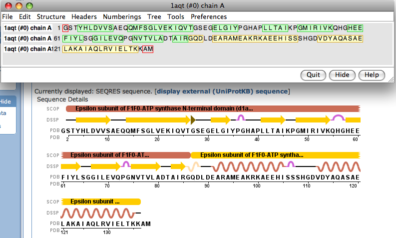
Sorry for the lack of details. I was referring the annotated sequence info for the PDB entry. Example that I mentioned: http://www.pdb.org/pdb/explore/remediatedSequence.do?structureId=1AQT It shows a DSSP cartoon of secondary structure above the sequence. I tried tweaking the parameters for ksdssp in Chimera (1.4) and could not get the same as shown with the PDB sequence. Cheers, Tom
I can only reiterate my first reply: <http://www.cgl.ucsf.edu/pipermail/chimera-users/2010-May/005149.html> Sorry I don't have anything more specific to tell you, Elaine On May 7, 2010, at 6:27 AM, Tom Duncan wrote:
On May 6, 2010, at 6:32 PM, Elaine Meng wrote:
At first I thought you meant you ran DSSP yourself. How do you know that the secondary structure in the PDB file of 1aqt came from DSSP? I thought that PDB structure depositors were at liberty to define secondary structure as they wished, which could be done with several different programs or even based on subjective opinions. In that case, it is not at all surprising for there to be differences, within reason. I could be mistaken about the PDB protocol, however.
Sorry for the lack of details. I was referring the annotated sequence info for the PDB entry. Example that I mentioned: http://www.pdb.org/pdb/explore/remediatedSequence.do?structureId=1AQT It shows a DSSP cartoon of secondary structure above the sequence. I tried tweaking the parameters for ksdssp in Chimera (1.4) and could not get the same as shown with the PDB sequence.

On May 7, 2010, at 6:27 AM, Tom Duncan wrote:
On May 6, 2010, at 6:32 PM, Elaine Meng wrote:
At first I thought you meant you ran DSSP yourself. How do you know that the secondary structure in the PDB file of 1aqt came from DSSP? I thought that PDB structure depositors were at liberty to define secondary structure as they wished, which could be done with several different programs or even based on subjective opinions. In that case, it is not at all surprising for there to be differences, within reason. I could be mistaken about the PDB protocol, however.
Sorry for the lack of details. I was referring the annotated sequence info for the PDB entry. Example that I mentioned: http://www.pdb.org/pdb/explore/remediatedSequence.do?structureId=1AQT It shows a DSSP cartoon of secondary structure above the sequence. I tried tweaking the parameters for ksdssp in Chimera (1.4) and could not get the same as shown with the PDB sequence.
I can't get them to be identical either, thought they're awfully close: The only difference I see is the tyrosine at position 35 (PDB numbering). Is this what you're referring to? Chimera has it as strand and the PDB does not. Looking at the structure it looks like the PDB might be correct in that the tyrosine doesn't seem to form any backbone hydrogen bonds with the neighboring strand. I'll have to look into this. --Eric Eric Pettersen UCSF Computer Graphics Lab http://www.cgl.ucsf.edu
participants (3)
-
 Elaine Meng
 -
 Eric Pettersen
 -
 Tom Duncan