ChimeraX version 1.9 has been released

UCSF ChimeraX version 1.9 has been released! ChimeraX includes user documentation and is free for noncommercial use. Download for Windows, Linux, and MacOS from: https://www.rbvi.ucsf.edu/chimerax/download.html Updates since version 1.8 (June 2024) include: - Foldseek search and analysis of large sets of similar structures - Find Cavities tool and "kvfinder" command to identify pockets and channels in macromolecules - optional residue-level summaries of H-bond and contact calculations - "mutationscores" command for analysis and plotting of results from deep mutational scanning - "surface hidefarblobs" command for colocalization analysis of light microscopy channels - raycasting option for medical images or other maps displayed as transparent volumes - selection can be expanded to all residues associated with the same column(s) in a sequence alignment - protein structure metadata can be updated with full chain sequences - missing-structure pseudobonds are labeled with the number of missing residues (default, can be turned off in preferences) For details, please see the ChimeraX change log: https://www.rbvi.ucsf.edu/trac/ChimeraX/wiki/ChangeLog On behalf of the team, Elaine ----- Elaine C. Meng, Ph.D. UCSF Chimera(X) team Resource for Biocomputing, Visualization, and Informatics Department of Pharmaceutical Chemistry University of California, San Francisco

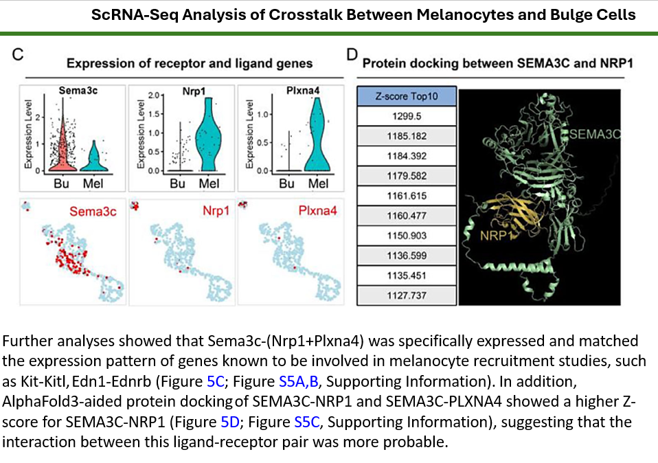
Dear Dr. Elaine Meng, Greetings, and Happy New Year. I hope you are doing well. I am writing to ask for your kind advice regarding a recent and excellent paper entitled “Regenerative Hair Pigmentation via Skin Organoids: Adaptive Patterning Mediated by Collagen VI and Semaphorin 3C.” In this study, the authors performed AlphaFold3-aided protein docking of SEMA3C with NRP1 and PLXNA4 and presented structural figures illustrating these interactions. I have attached the relevant figure ( at the end of this meesage) for your reference. May I kindly ask whether it is possible to generate similar structural visualization figures using UCSF ChimeraX? If so, I would greatly appreciate your guidance on whether ChimeraX can directly handle AlphaFold3-predicted complexes and produce comparable representations. Thank you very much for your time and kind consideration. I look forward to your advice. With best regards, =========================Ahmed Morsy, Ph.D.Assitant Professor, Department of Stem Cell & Regenerative Biotechnology, Konkuk University, 120 Neungdong-ro, Gwangjin-gu, Seoul05029, Korea C.P.: +82/10-5299-5844 E-mail: ahmed_morsy86@yahoo.com; ahmed@konkuk.ac.kr Orcidid: https://orcid.org/0000-0003-3873-9903 Scopus id: 55258308400 (https://www.scopus.com/authid/detail.uri?authorId=55258308400) Researchgate: https://www.researchgate.net/profile/Ahmed-Abdal-Dayem GoogleScholar: https://scholar.google.co.kr/citations?user=NevFBgMAAAAJ&hl=en On Saturday, December 14, 2024 at 02:35:50 AM GMT+9, Elaine Meng <meng@cgl.ucsf.edu> wrote: UCSF ChimeraX version 1.9 has been released! ChimeraX includes user documentation and is free for noncommercial use. Download for Windows, Linux, and MacOS from: https://www.rbvi.ucsf.edu/chimerax/download.html Updates since version 1.8 (June 2024) include: - Foldseek search and analysis of large sets of similar structures - Find Cavities tool and "kvfinder" command to identify pockets and channels in macromolecules - optional residue-level summaries of H-bond and contact calculations - "mutationscores" command for analysis and plotting of results from deep mutational scanning - "surface hidefarblobs" command for colocalization analysis of light microscopy channels - raycasting option for medical images or other maps displayed as transparent volumes - selection can be expanded to all residues associated with the same column(s) in a sequence alignment - protein structure metadata can be updated with full chain sequences - missing-structure pseudobonds are labeled with the number of missing residues (default, can be turned off in preferences) For details, please see the ChimeraX change log: https://www.rbvi.ucsf.edu/trac/ChimeraX/wiki/ChangeLog On behalf of the team, Elaine ----- Elaine C. Meng, Ph.D. UCSF Chimera(X) team Resource for Biocomputing, Visualization, and Informatics Department of Pharmaceutical Chemistry University of California, San Francisco _______________________________________________ ChimeraX-announce mailing list -- chimerax-announce@cgl.ucsf.edu To unsubscribe send an email to chimerax-announce-leave@cgl.ucsf.edu
Dear Ahmed Morsy, That is just showing the proteins as ribbons. Yes of course you can open protein structures and show them as ribbons. The labels of the protein names would need to be added separately, but you could do that with the 2D Labels tool or command. <https://rbvi.ucsf.edu/chimerax/docs/user/tools/2dlabels.html> See general information: making images with ChimeraX <https://rbvi.ucsf.edu/chimerax/docs/user/images.html> See also example images and command scripts: <https://www.rbvi.ucsf.edu/chimerax/gallery.html> <https://www.rbvi.ucsf.edu/chimerax/features.html> I hope this helps, Elaine ----- Elaine C. Meng, Ph.D. UCSF Chimera(X) team Resource for Biocomputing, Visualization, and Informatics Department of Pharmaceutical Chemistry University of California, San Francisco
On Jan 22, 2026, at 3:10 AM, ahmed morsy <ahmed_morsy86@yahoo.com> wrote:
Dear Dr. Elaine Meng, Greetings, and Happy New Year. I hope you are doing well. I am writing to ask for your kind advice regarding a recent and excellent paper entitled “Regenerative Hair Pigmentation via Skin Organoids: Adaptive Patterning Mediated by Collagen VI and Semaphorin 3C.” In this study, the authors performed AlphaFold3-aided protein docking of SEMA3C with NRP1 and PLXNA4 and presented structural figures illustrating these interactions. I have attached the relevant figure ( at the end of this meesage) for your reference. May I kindly ask whether it is possible to generate similar structural visualization figures using UCSF ChimeraX? If so, I would greatly appreciate your guidance on whether ChimeraX can directly handle AlphaFold3-predicted complexes and produce comparable representations. Thank you very much for your time and kind consideration. I look forward to your advice. With best regards, ========================= Ahmed Morsy, Ph.D. Assitant Professor,
Department of Stem Cell & Regenerative Biotechnology, Konkuk University, 120 Neungdong-ro, Gwangjin-gu, Seoul 05029, Korea C.P.: +82/10-5299-5844 E-mail: ahmed_morsy86@yahoo.com; ahmed@konkuk.ac.kr Orcid id: https://orcid.org/0000-0003-3873-9903 Scopus id: 55258308400 (https://www.scopus.com/authid/detail.uri?authorId=55258308400) Researchgate: https://www.researchgate.net/profile/Ahmed-Abdal-Dayem GoogleScholar: https://scholar.google.co.kr/citations?user=NevFBgMAAAAJ&hl=en <1769080147269blob.jpg>

Dear Dr. Morsey, Here are instructions on how to open AlphaFold3-generated structures in ChimeraX (I think this may have been the gist of your question?): 1. If you run a job on the AlphaFold server, you can download the results as a .zip file. 2. If you unzip it, you will see 15 files, 5 of which are .cif files. There are 5 .cif files because for each job, AF3 produces five predicted structures by sampling the diffusion process five times. The fold_(your job name)_model0.cif file is the one with the best metrics (ipTM, pTM) 3. You can open that .cif file in ChimeraX and work with it from there. I hope this was useful. I will also point you to our recent preprint here <https://www.biorxiv.org/content/10.64898/2026.01.06.698042v1> where we used AF3 to predict novel GLI-SUFU structures and ChimeraX to visualize/analyze them. _________________________________ Lee Bardwell, Ph.D. Professor and Vice-Chair, Dept. of Developmental & Cell Biology Dunlop School of Biological Sciences University of California, Irvine https://faculty.sites.uci.edu/bardwell/ __________________________________ On Jan 22, 2026, at 8:36 AM, Elaine Meng via ChimeraX-users <chimerax-users@cgl.ucsf.edu> wrote: Dear Ahmed Morsy, That is just showing the proteins as ribbons. Yes of course you can open protein structures and show them as ribbons. The labels of the protein names would need to be added separately, but you could do that with the 2D Labels tool or command. <https://urldefense.com/v3/__https://rbvi.ucsf.edu/chimerax/docs/user/tools/2... > See general information: making images with ChimeraX <https://urldefense.com/v3/__https://rbvi.ucsf.edu/chimerax/docs/user/images.... > See also example images and command scripts: <https://urldefense.com/v3/__https://www.rbvi.ucsf.edu/chimerax/gallery.html_... > <https://urldefense.com/v3/__https://www.rbvi.ucsf.edu/chimerax/features.html... > I hope this helps, Elaine ----- Elaine C. Meng, Ph.D. UCSF Chimera(X) team Resource for Biocomputing, Visualization, and Informatics Department of Pharmaceutical Chemistry University of California, San Francisco On Jan 22, 2026, at 3:10 AM, ahmed morsy <ahmed_morsy86@yahoo.com<mailto:ahmed_morsy86@yahoo.com>> wrote: Dear Dr. Elaine Meng, Greetings, and Happy New Year. I hope you are doing well. I am writing to ask for your kind advice regarding a recent and excellent paper entitled “Regenerative Hair Pigmentation via Skin Organoids: Adaptive Patterning Mediated by Collagen VI and Semaphorin 3C.” In this study, the authors performed AlphaFold3-aided protein docking of SEMA3C with NRP1 and PLXNA4 and presented structural figures illustrating these interactions. I have attached the relevant figure ( at the end of this meesage) for your reference. May I kindly ask whether it is possible to generate similar structural visualization figures using UCSF ChimeraX? If so, I would greatly appreciate your guidance on whether ChimeraX can directly handle AlphaFold3-predicted complexes and produce comparable representations. Thank you very much for your time and kind consideration. I look forward to your advice. With best regards, ========================= Ahmed Morsy, Ph.D. Assitant Professor, Department of Stem Cell & Regenerative Biotechnology, Konkuk University, 120 Neungdong-ro, Gwangjin-gu, Seoul 05029, Korea C.P.: +82/10-5299-5844 E-mail: ahmed_morsy86@yahoo.com; ahmed@konkuk.ac.kr Orcid id: https://urldefense.com/v3/__https://orcid.org/0000-0003-3873-9903__;!!CzAuKJ... Scopus id: 55258308400 (https://urldefense.com/v3/__https://www.scopus.com/authid/detail.uri?authorI... ) Researchgate: https://urldefense.com/v3/__https://www.researchgate.net/profile/Ahmed-Abdal... GoogleScholar: https://urldefense.com/v3/__https://scholar.google.co.kr/citations?user=NevF... <1769080147269blob.jpg> _______________________________________________ ChimeraX-users mailing list -- chimerax-users@cgl.ucsf.edu<mailto:chimerax-users@cgl.ucsf.edu> To unsubscribe send an email to chimerax-users-leave@cgl.ucsf.edu<mailto:chimerax-users-leave@cgl.ucsf.edu> Archives: https://urldefense.com/v3/__https://mail.cgl.ucsf.edu/mailman/archives/list/...
Dear Professor Bardwell, Thank you very much for the expalanation and sharring your recent related publication. Very appreciated! Have a nice day, Best regards, Ahmed Morsy ========================= Ahmed Morsy, Ph.D. Assitant Professor, Department of Stem Cell & Regenerative Biotechnology, Konkuk University, 120 Neungdong-ro, Gwangjin-gu, Seoul 05029, Korea C.P.: +82/10-5299-5844 E-mail: ahmed_morsy86@yahoo.com; ahmed@konkuk.ac.kr Orcid id: https://urldefense.com/v3/__https://orcid.org/0000-0003-3873-9903__;!!CzAuKJ... Scopus id: 55258308400 (https://urldefense.com/v3/__https://www.scopus.com/authid/detail.uri?authorI... ) Researchgate: https://urldefense.com/v3/__https://www.researchgate.net/profile/Ahmed-Abdal... GoogleScholar: https://urldefense.com/v3/__https://scholar.google.co.kr/citations?user=NevF... <1769080147269blob.jpg> On Friday, January 23, 2026 at 03:10:52 AM GMT+9, Lee Bardwell <bardwell@uci.edu> wrote: Dear Dr. Morsey, Here are instructions on how to open AlphaFold3-generated structures in ChimeraX (I think this may have been the gist of your question?): 1. If you run a job on the AlphaFold server, you can download the results as a .zip file. 2. If you unzip it, you will see 15 files, 5 of which are .cif files. There are 5 .cif files because for each job, AF3 produces five predicted structures by sampling the diffusion process five times.The fold_(your job name)_model0.cif file is the one with the best metrics (ipTM, pTM) 3. You can open that .cif file in ChimeraX and work with it from there. I hope this was useful. I will also point you to our recent preprint here where we used AF3 to predict novel GLI-SUFU structures and ChimeraX to visualize/analyze them._________________________________ Lee Bardwell, Ph.D. Professor and Vice-Chair, Dept. of Developmental & Cell Biology Dunlop School of Biological Sciences University of California, Irvinehttps://faculty.sites.uci.edu/bardwell/ __________________________________ On Jan 22, 2026, at 8:36 AM, Elaine Meng via ChimeraX-users <chimerax-users@cgl.ucsf.edu> wrote: Dear Ahmed Morsy, That is just showing the proteins as ribbons. Yes of course you can open protein structures and show them as ribbons. The labels of the protein names would need to be added separately, but you could do that with the 2D Labels tool or command. <https://urldefense.com/v3/__https://rbvi.ucsf.edu/chimerax/docs/user/tools/2... > See general information: making images with ChimeraX <https://urldefense.com/v3/__https://rbvi.ucsf.edu/chimerax/docs/user/images.... > See also example images and command scripts: <https://urldefense.com/v3/__https://www.rbvi.ucsf.edu/chimerax/gallery.html_... > <https://urldefense.com/v3/__https://www.rbvi.ucsf.edu/chimerax/features.html... > I hope this helps, Elaine ----- Elaine C. Meng, Ph.D. UCSF Chimera(X) team Resource for Biocomputing, Visualization, and Informatics Department of Pharmaceutical Chemistry University of California, San Francisco On Jan 22, 2026, at 3:10 AM, ahmed morsy <ahmed_morsy86@yahoo.com> wrote: Dear Dr. Elaine Meng, Greetings, and Happy New Year. I hope you are doing well. I am writing to ask for your kind advice regarding a recent and excellent paper entitled “Regenerative Hair Pigmentation via Skin Organoids: Adaptive Patterning Mediated by Collagen VI and Semaphorin 3C.” In this study, the authors performed AlphaFold3-aided protein docking of SEMA3C with NRP1 and PLXNA4 and presented structural figures illustrating these interactions. I have attached the relevant figure ( at the end of this meesage) for your reference. May I kindly ask whether it is possible to generate similar structural visualization figures using UCSF ChimeraX? If so, I would greatly appreciate your guidance on whether ChimeraX can directly handle AlphaFold3-predicted complexes and produce comparable representations. Thank you very much for your time and kind consideration. I look forward to your advice. With best regards, ========================= Ahmed Morsy, Ph.D. Assitant Professor, Department of Stem Cell & Regenerative Biotechnology, Konkuk University, 120 Neungdong-ro, Gwangjin-gu, Seoul 05029, Korea C.P.: +82/10-5299-5844 E-mail: ahmed_morsy86@yahoo.com; ahmed@konkuk.ac.kr Orcid id: https://urldefense.com/v3/__https://orcid.org/0000-0003-3873-9903__;!!CzAuKJ... Scopus id: 55258308400 (https://urldefense.com/v3/__https://www.scopus.com/authid/detail.uri?authorI... ) Researchgate: https://urldefense.com/v3/__https://www.researchgate.net/profile/Ahmed-Abdal... GoogleScholar: https://urldefense.com/v3/__https://scholar.google.co.kr/citations?user=NevF... <1769080147269blob.jpg> _______________________________________________ ChimeraX-users mailing list -- chimerax-users@cgl.ucsf.edu To unsubscribe send an email to chimerax-users-leave@cgl.ucsf.edu Archives: https://urldefense.com/v3/__https://mail.cgl.ucsf.edu/mailman/archives/list/...
participants (3)
-
 ahmed morsy
 -
 Elaine Meng
 -
 Lee Bardwell