Fwd: opening cryoEM SPA maps using "volume" view mode

Begin forwarded message:
From: Jonathan Paul Remis <jpremis@berkeley.edu> Subject: opening cryoEM SPA maps using "volume" view mode Date: May 18, 2026 at 4:36:10 PM PDT To: chimerax-bugs@cgl.ucsf.edu
Hello wonderful people that build and maintain Chimera!
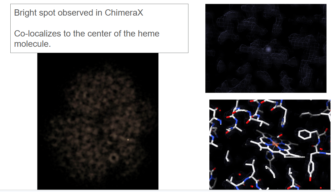
I have run into a bit of an odd observation which when compounded with some basic biochemistry has left me scratching my head. Essentially, when I open one of our SPA volumes in chimerax and choos "volume" in the volume viewer I am able to see a single bright spot in the volume. If I process our data with C2 symmetry we see two bright spots.
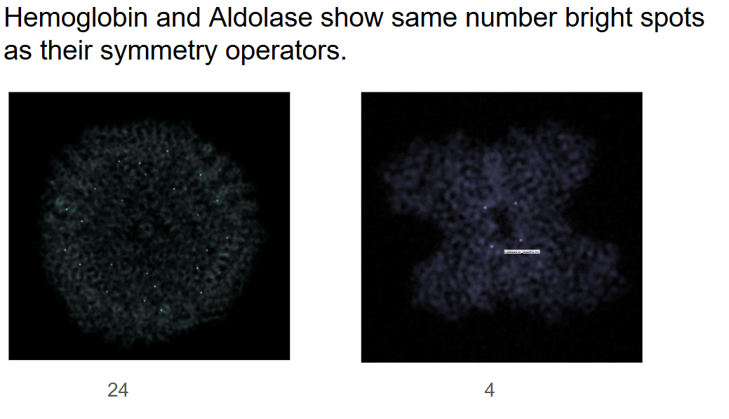
I had thought this was biologically relevant but I opened up a couple more structures from our lab and other labs and see this is pretty common. Please see the attached images and you can see ApoFerritin has 24 bright spots, and Aldolase has 4.
In the case of my sample - hemoglobin - the bright spot co-localizes with a iron element in the structure. I want to believe this has something to do with the density/scattering of the Fe but am at a loss for why we would not see 4 of them. Hemoglobin has 4 heme centers and 4 Fe atoms.
Thank you in advance for any suggestions or support you can provide.
Thanks, Remis


-- Jonathan Remis Scientific Engineer Müller Lab University of California, Berkeley

Hello Remis, The "volume" type of display is just a transparent blob where higher densities are rendered as higher intensities. So all I can say is that the dot(s) are the highest value(s) in your data. I.e. potentially (no pun intended) resulting from the actual atomic structure, unless there is some kind of experimental artifact that would put a spurious peak in the map. Maybe there are 4 peaks for your 4 ions but due to the particle averaging process, conformational heterogeneity, etc. one of them is higher than the others? Maybe to get a better understanding of what's there, you could (1) adjust the transfer function by dragging the threshold positions and heights on the histogram in Volume Viewer, and/or creating more thresholds. The default initial positions are not generally the best for understanding your specific data. Alternatively you may want to try #2 below (2) show the data as "surface" or "mesh" (isosurfaces) instead of as "volume." The isosurfaces are conceptually simpler, whereas volume transparency/brightness isn't always completely intuitive (to me, anyway). Then you can adjust the isosurface threshold (contour) level by dragging the vertical bar left/right on the histogram. See the Volume Viewer help: <https://rbvi.ucsf.edu/chimerax/docs/user/tools/volumeviewer.html> I hope this helps, Elaine ----- Elaine C. Meng, Ph.D. UCSF Chimera(X) team Resource for Biocomputing, Visualization, and Informatics Department of Pharmaceutical Chemistry University of California, San Francisco
On May 19, 2026, at 12:54 PM, Eric Pettersen via ChimeraX-users <chimerax-users@cgl.ucsf.edu> wrote:
Begin forwarded message:
From: Jonathan Paul Remis <jpremis@berkeley.edu> Subject: opening cryoEM SPA maps using "volume" view mode Date: May 18, 2026 at 4:36:10 PM PDT To: chimerax-bugs@cgl.ucsf.edu
Hello wonderful people that build and maintain Chimera!
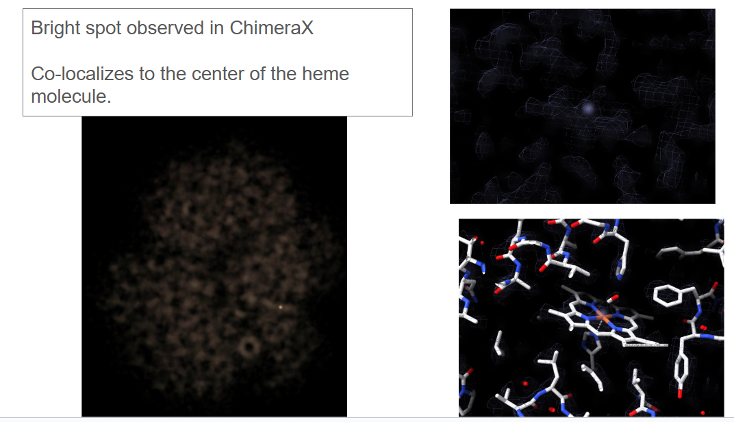
I have run into a bit of an odd observation which when compounded with some basic biochemistry has left me scratching my head. Essentially, when I open one of our SPA volumes in chimerax and choos "volume" in the volume viewer I am able to see a single bright spot in the volume. If I process our data with C2 symmetry we see two bright spots.
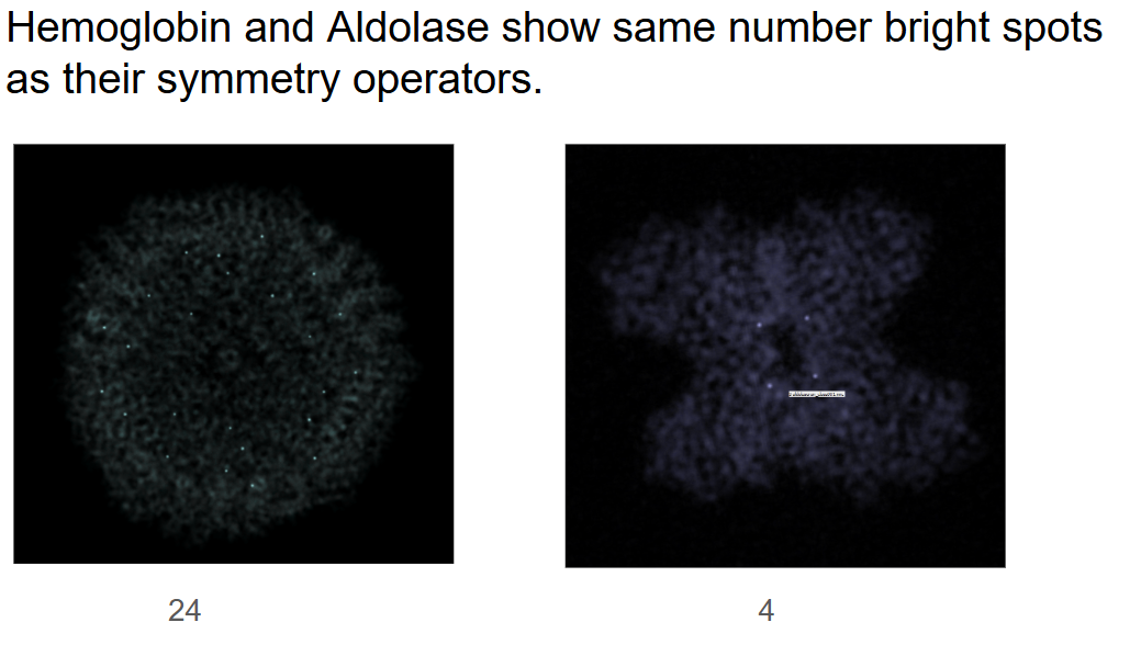
I had thought this was biologically relevant but I opened up a couple more structures from our lab and other labs and see this is pretty common. Please see the attached images and you can see ApoFerritin has 24 bright spots, and Aldolase has 4.
In the case of my sample - hemoglobin - the bright spot co-localizes with a iron element in the structure. I want to believe this has something to do with the density/scattering of the Fe but am at a loss for why we would not see 4 of them. Hemoglobin has 4 heme centers and 4 Fe atoms.
Thank you in advance for any suggestions or support you can provide.
Thanks, Remis
-- Jonathan Remis Scientific Engineer Müller Lab University of California, Berkeley
_______________________________________________ ChimeraX-users mailing list -- chimerax-users@cgl.ucsf.edu To unsubscribe send an email to chimerax-users-leave@cgl.ucsf.edu Archives: https://mail.cgl.ucsf.edu/mailman/archives/list/chimerax-users@cgl.ucsf.edu/
participants (2)
-
 Elaine Meng
 -
 Eric Pettersen