A question for GPI-anchored protein

Dear DR. Thank you for using chimeraX. I am very impressed with the development of protein assembly analysis technology. I am an elderly (88-year-old) biochemist. I have a question. ○1. Even if dimer formation is analyzed by AlphaFold 3, dimers with interfaces are not recognized at all (AlphaFold has a list of 19(?) analyzed). Why? ○2. Even though dimer formation (54 or 132 interface residues) can be seen by chimeraX, when looking at ipTM with AlphaFold, a number such as 0.19 is not recognized as a dimer. Do you think that this is because AlphaFold does not take into account that proteins such as GPI membrane proteins are kept in anti-parallel, do you have an opinion? With best Regards Hiroshi Ochiai Research Institute for Electronic Sciences, Hokkaido University, Sapporo Japan Emeritus Professor
Dear Dr, I forgot to add the comment that the protein forms dimer on the SDS-PAGE. “Dimer Formation of a Cell-Cell Adhesion Protein, gp64 of the Cellular Slime Mold, Polysphondylium pallidum” Plans and Cell Physiology, Vol. 36, March 1996, pages 135-139 Hiroshi
2026/07/05 14:29、落合廣 <ochiai3@icloud.com>のメール:
Dear DR. Thank you for using chimeraX. I am very impressed with the development of protein assembly analysis technology. I am an elderly (88-year-old) biochemist. I have a question. ○1. Even if dimer formation is analyzed by AlphaFold 3, dimers with interfaces are not recognized at all (AlphaFold has a list of 19(?) analyzed). Why? ○2. Even though dimer formation (54 or 132 interface residues) can be seen by chimeraX, when looking at ipTM with AlphaFold, a number such as 0.19 is not recognized as a dimer. Do you think that this is because AlphaFold does not take into account that proteins such as GPI membrane proteins are kept in anti-parallel, do you have an opinion? With best Regards
Hiroshi Ochiai Research Institute for Electronic Sciences, Hokkaido University, Sapporo Japan Emeritus Professor

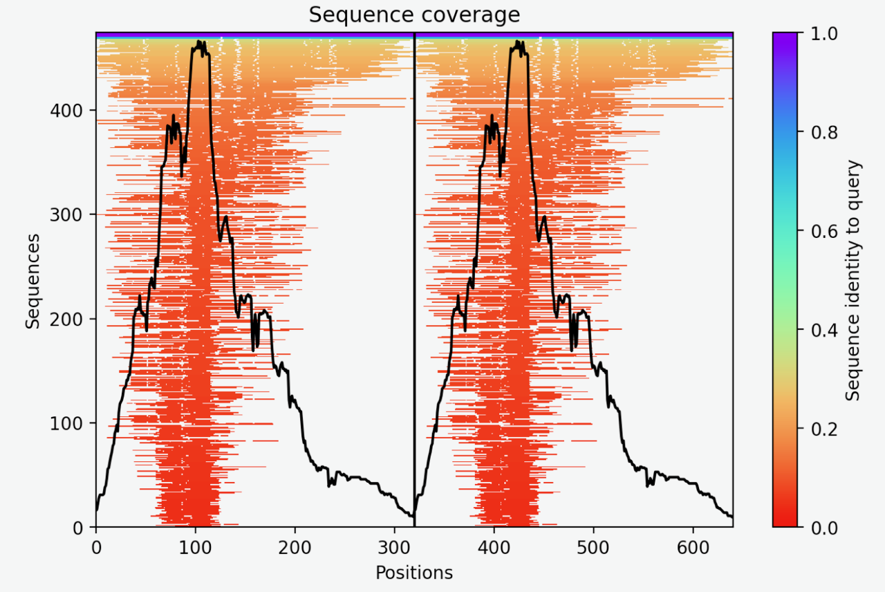
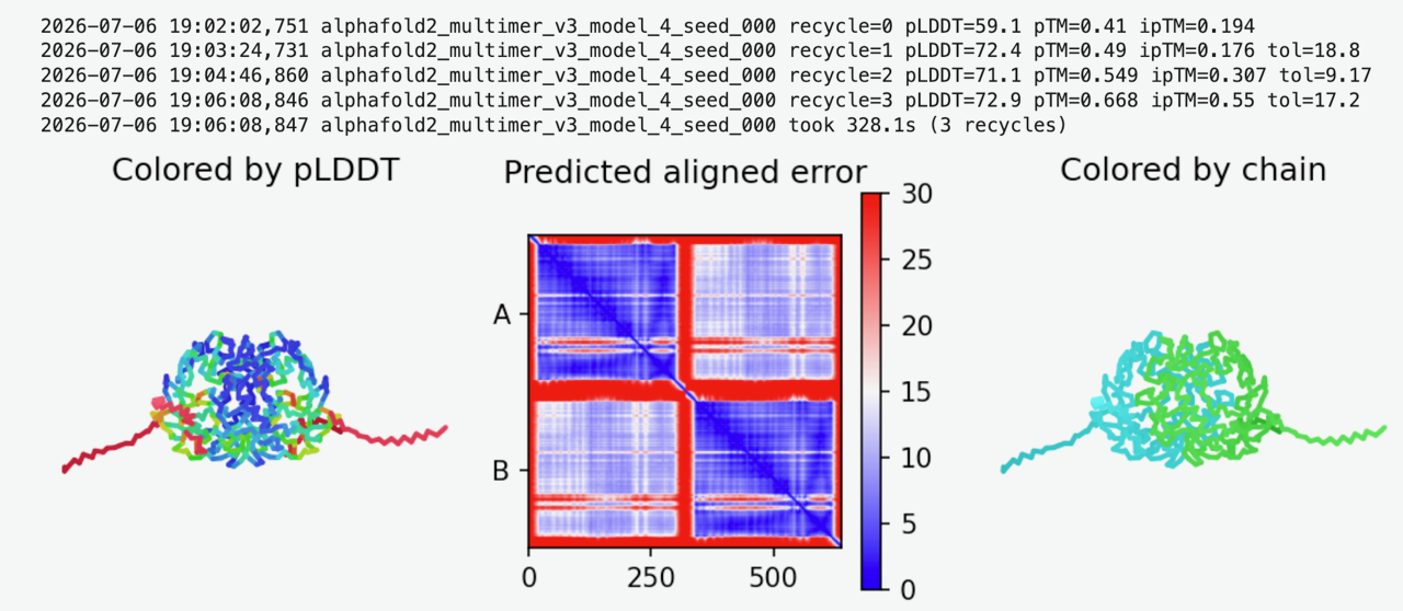
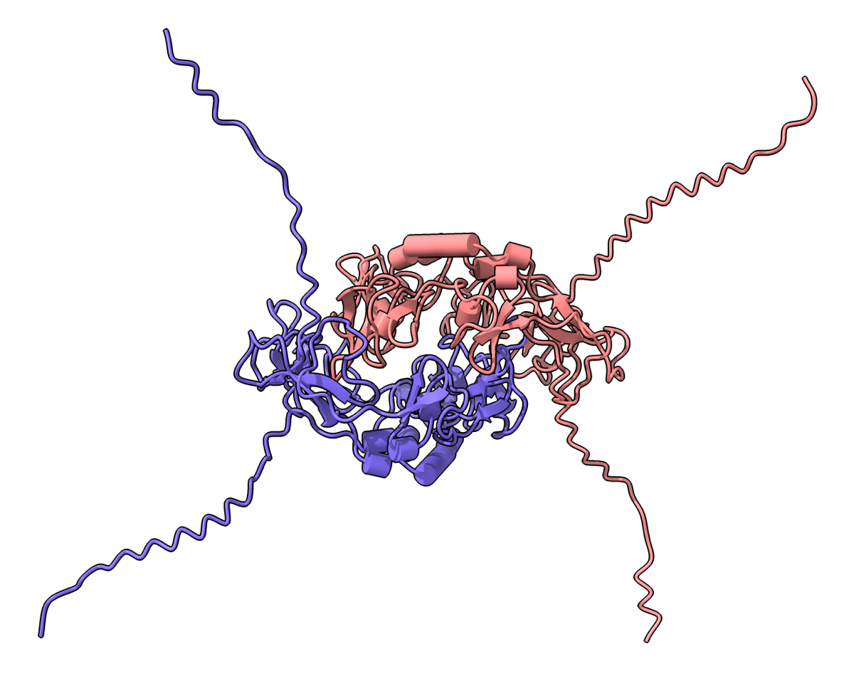
Hi Hiroshi, Machine learning structure prediction programs such as AlphaFold 3, AlphaFold 2, Boltz 2, and OpenFold 3 are not always able to correctly predict protein-protein interactions. These methods all use a multiple sequence alignments (MSA) computed from similar sequences from databases containing as many as a few billion experimentally observed sequences. If there are few known sequences similar to the one you are predicting this can result in poor quality predictions. In the case of your cell-cell adhesion protein gp64 from Polysphondylium pallidum (UniProt Q52085) there are very few similar sequences, only a few hundred. High quality predictions usually have MSAs containing thousands of sequences. ChimeraX can run predictions using AlphaFold 2 (menu Tools / Structure Prediction / AlphaFold), or Boltz 2, or OpenFold 3. I ran your dimer with all 3 of these methods and also with Google's AlphaFold 3 server. With Boltz 2, OpenFold 3, and AlphaFold 3 I got very poor interface predicted TM scores (ipTM) of 0.19, 0.16, and 0.23 similar to what you report. This indicates those programs have no confidence that the dimer interface is correct. Interestingly AlphaFold 2 (run using ChimeraX on Google Colab), which is by far the oldest of these 4 prediction programs (July 2021) gave an ipTM score of 0.55. That is still not a very confident score. Confident scores would be around 0.8 - 1.0. But it might be worth looking at the AlphaFold 2 predictions. I've attached the AlphaFold 2 prediction results (results.zip) below. Tom Here is the AlphaFold 2 sequence coverage plot showing it used an MSA with about 450 similar sequences, although only about 5 had high similarity (blue color).  Here is the AlphaFold 2 best prediction out of 5. The predicted aligned error plot off-diagonal blocks indicate some confidence in the dimer interface.  Here is an image of the dimer in ChimeraX  
On Jul 4, 2026, at 10:29 PM, 落合廣 via ChimeraX-users <chimerax-users@cgl.ucsf.edu> wrote:
Dear DR. Thank you for using chimeraX. I am very impressed with the development of protein assembly analysis technology. I am an elderly (88-year-old) biochemist. I have a question. ○1. Even if dimer formation is analyzed by AlphaFold 3, dimers with interfaces are not recognized at all (AlphaFold has a list of 19(?) analyzed). Why? ○2. Even though dimer formation (54 or 132 interface residues) can be seen by chimeraX, when looking at ipTM with AlphaFold, a number such as 0.19 is not recognized as a dimer. Do you think that this is because AlphaFold does not take into account that proteins such as GPI membrane proteins are kept in anti-parallel, do you have an opinion? With best Regards
Hiroshi Ochiai Research Institute for Electronic Sciences, Hokkaido University, Sapporo Japan Emeritus Professor _______________________________________________ ChimeraX-users mailing list -- chimerax-users@cgl.ucsf.edu To unsubscribe send an email to chimerax-users-leave@cgl.ucsf.edu Archives: https://mail.cgl.ucsf.edu/mailman/archives/list/chimerax-users@cgl.ucsf.edu/
HI DR, I lost your mail but not your replayed data Thank you very much for your quick responses and for your efforts in analyzing our cell-cell adhesion protein, gp64. As I am a beginner in this field, I might not always be able to reply to you quickly. However, I will get back to you with more questions soon. thak you again Hiroshi
2026/07/05 17:16、落合廣 <ochiai3@icloud.com>のメール:
Dear Dr,
I forgot to add the comment that the protein forms dimer on the SDS-PAGE.
“Dimer Formation of a Cell-Cell Adhesion Protein, gp64 of the Cellular Slime Mold, Polysphondylium pallidum” Plans and Cell Physiology, Vol. 36, March 1996, pages 135-139
Hiroshi
2026/07/05 14:29、落合廣 <ochiai3@icloud.com>のメール:
Dear DR. Thank you for using chimeraX. I am very impressed with the development of protein assembly analysis technology. I am an elderly (88-year-old) biochemist. I have a question. ○1. Even if dimer formation is analyzed by AlphaFold 3, dimers with interfaces are not recognized at all (AlphaFold has a list of 19(?) analyzed). Why? ○2. Even though dimer formation (54 or 132 interface residues) can be seen by chimeraX, when looking at ipTM with AlphaFold, a number such as 0.19 is not recognized as a dimer. Do you think that this is because AlphaFold does not take into account that proteins such as GPI membrane proteins are kept in anti-parallel, do you have an opinion? With best Regards
Hiroshi Ochiai Research Institute for Electronic Sciences, Hokkaido University, Sapporo Japan Emeritus Professor
Hi Dr, Thank you very much for conducting the analysis using various versions of AlphaFold. Based on the AlphaFold criteria, the conclusion appears to be that gp64 is unlikely to form a proper dimer. While I may have some misunderstandings due to my limited expertise in using AlphaFold and ChimeraX, I believe that—considering the following points—it cannot be definitively ruled out that gp64 forms a dimer. Specifically, AlphaFold3 predicts the structure as a "protein moving freely in solution," whereas in reality, gp64 is anchored to the membrane surface via a GPI anchor. 1. A dimer-like band is observed in SDS-PAGE. 2. It contains a Sushi domain (characterized by four disulfide bridges); Sushi domains are known to be involved in protein-protein associations (as seen in "complement-containing proteins"). 3. ChimeraX analysis indicates an interface involving approximately 58 residues. 4. The interface surface area is 1,398 Ų. Given these factors, I would appreciate your opinion on how this should be interpreted. with best regards, Hiroshi Ochiai
2026/07/05 14:29、落合廣 <ochiai3@icloud.com>のメール:
Dear DR. Thank you for using chimeraX. I am very impressed with the development of protein assembly analysis technology. I am an elderly (88-year-old) biochemist. I have a question. ○1. Even if dimer formation is analyzed by AlphaFold 3, dimers with interfaces are not recognized at all (AlphaFold has a list of 19(?) analyzed). Why? ○2. Even though dimer formation (54 or 132 interface residues) can be seen by chimeraX, when looking at ipTM with AlphaFold, a number such as 0.19 is not recognized as a dimer. Do you think that this is because AlphaFold does not take into account that proteins such as GPI membrane proteins are kept in anti-parallel, do you have an opinion? With best Regards
Hiroshi Ochiai Research Institute for Electronic Sciences, Hokkaido University, Sapporo Japan Emeritus Professor
Hi Hiroshi, I think everyone agrees that you cannot trust AlphaFold or other computational structure prediction methods to decide if proteins bind. These computational predictions are just to give you clues about the possible behavior and must be tested experimentally. In the case of gp64 AlphaFold 3, Boltz 2, OpenFold 3, and AlphaFold 2 all have low confidence that it forms a dimer. But if experiments show it forms a dimer then quite possibly the computational predictions are wrong. Of course you have to just the weight and reliability of experimental evidence also. I am not an experimentalist and do not know how reliable an SDS-PAGE gel is at identifying oligomeric protein states. The computational predictions are not for "proteins moving freely in solution". These computational programs know nothing about water or dynamics of proteins. They were trained on experimental structures from the Protein DataBank which are mostly derived from X-ray crystallography and electron cryo-microscopy, and those techniques have the proteins frozen in non-crystalline ice. Also as you point out gp64 is attached to membranes, and the membrane environment may be necessary for them to dimerize. In summary, the computational predictions do not provide decisive answers about protein-protein interactions. Tom
On Jul 8, 2026, at 5:25 PM, 落合廣 via ChimeraX-users <chimerax-users@cgl.ucsf.edu> wrote:
Hi Dr, Thank you very much for conducting the analysis using various versions of AlphaFold. Based on the AlphaFold criteria, the conclusion appears to be that gp64 is unlikely to form a proper dimer. While I may have some misunderstandings due to my limited expertise in using AlphaFold and ChimeraX, I believe that—considering the following points—it cannot be definitively ruled out that gp64 forms a dimer. Specifically, AlphaFold3 predicts the structure as a "protein moving freely in solution," whereas in reality, gp64 is anchored to the membrane surface via a GPI anchor. 1. A dimer-like band is observed in SDS-PAGE. 2. It contains a Sushi domain (characterized by four disulfide bridges); Sushi domains are known to be involved in protein-protein associations (as seen in "complement-containing proteins"). 3. ChimeraX analysis indicates an interface involving approximately 58 residues. 4. The interface surface area is 1,398 Ų. Given these factors, I would appreciate your opinion on how this should be interpreted. with best regards,
Hiroshi Ochiai
2026/07/05 14:29、落合廣 <ochiai3@icloud.com>のメール:
Dear DR. Thank you for using chimeraX. I am very impressed with the development of protein assembly analysis technology. I am an elderly (88-year-old) biochemist. I have a question. ○1. Even if dimer formation is analyzed by AlphaFold 3, dimers with interfaces are not recognized at all (AlphaFold has a list of 19(?) analyzed). Why? ○2. Even though dimer formation (54 or 132 interface residues) can be seen by chimeraX, when looking at ipTM with AlphaFold, a number such as 0.19 is not recognized as a dimer. Do you think that this is because AlphaFold does not take into account that proteins such as GPI membrane proteins are kept in anti-parallel, do you have an opinion? With best Regards
Hiroshi Ochiai Research Institute for Electronic Sciences, Hokkaido University, Sapporo Japan Emeritus Professor
_______________________________________________ ChimeraX-users mailing list -- chimerax-users@cgl.ucsf.edu To unsubscribe send an email to chimerax-users-leave@cgl.ucsf.edu Archives: https://mail.cgl.ucsf.edu/mailman/archives/list/chimerax-users@cgl.ucsf.edu/
participants (2)
-
 Tom Goddard
 -
 落合廣
落合廣